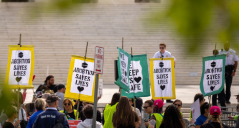
Anti-abortion rhetoric and images saturating the internet and political discussions portray embryos in early pregnancy as fully formed miniature human beings. Recently enacted abortion bans refer to embryos as “babies” and claim there’s a “fetal heartbeat” at six weeks of pregnancy. But what does an early pregnancy actually look like?
The MYA Network (short for “My Abortion Network”) set out to answer this question in their recent Issue of Tissue Project, which shows what tissue removed during early abortions looks like. The MYA Network is composed of clinicians working to expand early abortion options in primary care settings using abortion pills and manual uterine aspiration.
“There’s a lot of misinformation out there and many people have come to believe it,” said MYA Network co-founder Dr. Michele Gomez, a family care physician in Burlingame, Calif. “There’s also accurate information out there but mostly in the form of highly magnified embryos at these early stages. People have a right to know and understand that they are magnified.”
In fact, before nine weeks of pregnancy—when 80 percent of abortions take place—the embryo is not visible with the naked eye in the pregnancy tissue removed during an abortion. There is no “heartbeat” at six weeks of pregnancy, only the electrical activity of cells before an actual heart is formed, said Gomez.
“The anti-choice movement has been showing inaccurate and graphic pictures for a long time, and these images shock us so they tend to stick with us,” said Gomez. “Without factual images to counter the inaccurate images, many people just accept the only things they’ve ever seen, which is completely understandable. Seeing actual images of early pregnancy tissue, as seen with the naked eye, can help people replace the graphic and inaccurate images they may have come to accept as true.”
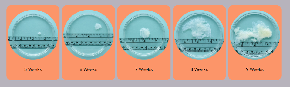
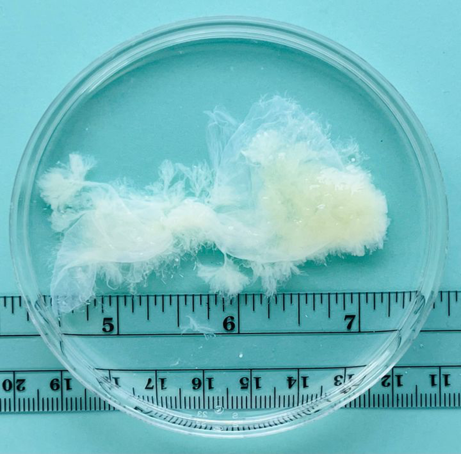
MYA Network created the Issue of Tissue Project to share accurate images of early pregnancy tissue. After manual aspiration abortions, they rinsed the blood from the tissue removed from the uterus and photographed it for pregnancies of five weeks through nine weeks.
Seeing actual images of early pregnancy tissue, as seen with the naked eye, can help people replace the graphic and inaccurate images they may have come to accept as true.
Dr. Michele Gomez
After an egg joins with a sperm at fertilization, it’s called a zygote. After five days of development, it’s called a blastocyst. In week four of the pregnancy, the blastocyst embeds in the uterine wall and becomes an embryo. At around the 10th week of pregnancy it becomes a fetus. Up to 50 percent of all pregnancies end in miscarriage.
In the MYA Network pregnancy tissue photos, the embryos are too small to see with the naked eye. At five weeks of pregnancy, the tissue is about one quarter of an inch wide.
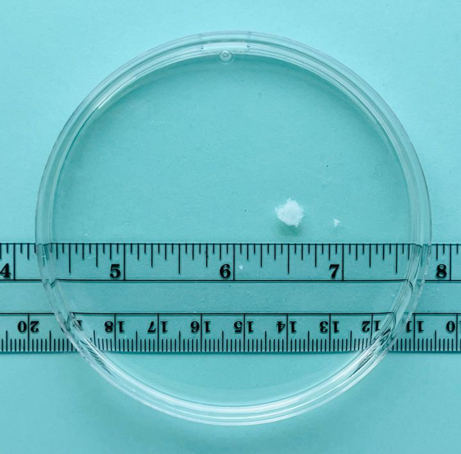
At six weeks of pregnancy, the tissue is a little over half an inch.
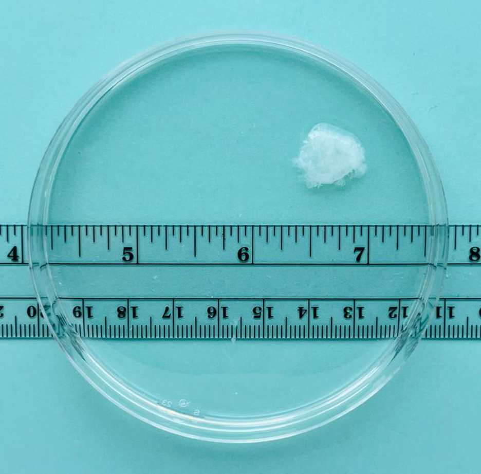
At seven weeks of pregnancy, the tissue is one inch wide.
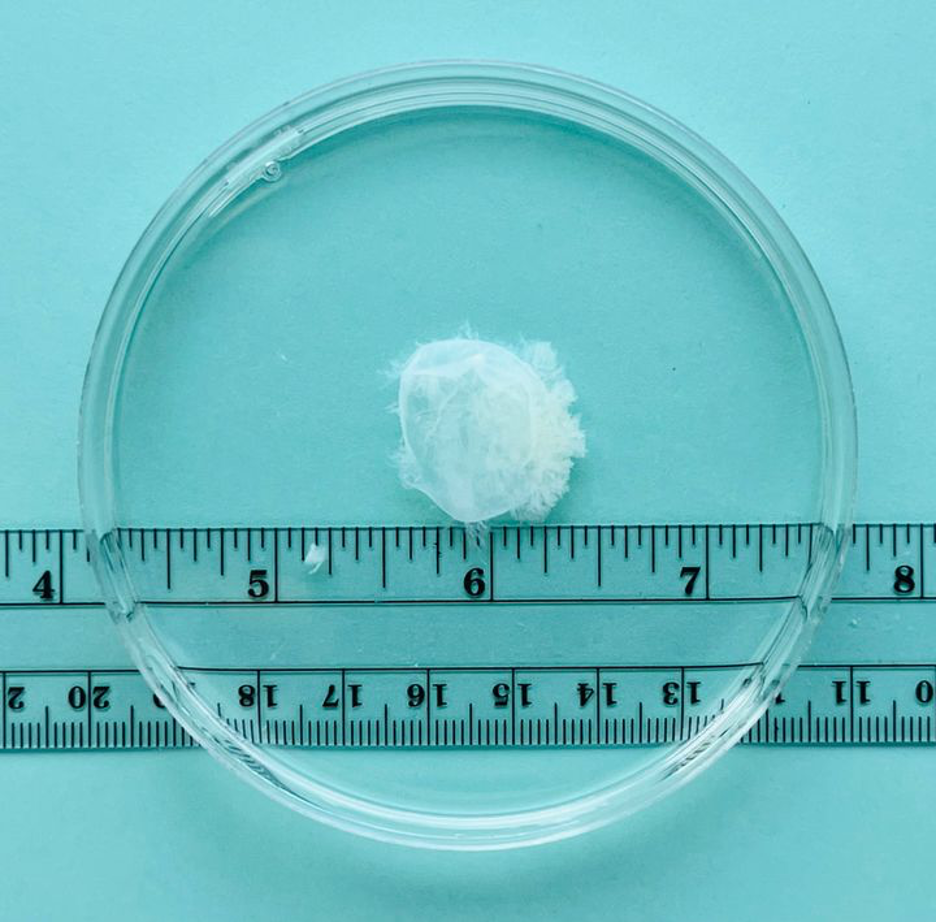
At eight weeks of pregnancy, the tissue is two and a half inches wide.
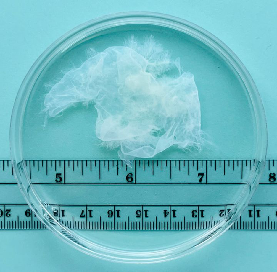
At nine weeks of pregnancy, the tissue is about three inches wide.
Gomez said the MYA Network’s decision to share these images grew from their own experiences as clinicians.
“Many of us in the MYA Network provide in-office abortions by manual uterine aspiration, which a simple, non-surgical procedure that takes five to 10 minutes to complete and can be done with only ibuprofen for pain control,” she said. “We’ve often had the experience of a patient asking to see the pregnancy tissue after the procedure, and then being very surprised by what they see.
“In my experience, pregnant people and their partners have felt some relief when they see the tissue, because it doesn’t look like what they’ve seen in anti-choice imagery,” Gomez continued. “We in the MYA Network discussed this, and decided that as clinicians we wanted our patients to have more information, so they could be better informed.”
Seeing the tissue resulting from pregnancy termination can dispel the myths surrounding abortion, help clear up confusion and misunderstandings resulting from anti-abortion misrepresentations, and combat abortion stigma and shame.
On their website, the MYA Network shares recordings of patients speaking about their surprise and relief after viewing their own pregnancy tissue after an abortion.
One woman explained, “It wasn’t what I expected at all. It was so small … and seemed a lot less scary.”
Others spoke about how seeing their pregnancy tissue relieved feelings of guilt. “It just looked like mucus, it was just a little thing … nothing that made us feel guilty,” said another patient after viewing her pregnancy tissue.
Explaining why MYA Network decided to share these patients’ reactions, Gomez said, “We wanted to make the information more personal—facts are helpful, but there are so many feelings that can come up with viewing these images, and everyone’s feelings are valid. Our website is visited mostly by people who may be considering abortion, and these people often feel alone—they may not feel comfortable talking to many people, or even to any person about their situation. But humans are social beings, so hearing other peoples’ experiences can be helpful when we need to make decisions about our own lives.”
TikTok influencers are now using the MYA Network images to raise awareness about early abortion and point out the absurdity of this early pregnancy tissue having more rights than the actual person who is pregnant.
@auntiekilljoy #greenscreen they need to show these images at every political debate about abortion #feminism #abortion #fyp ♬ original sound – Jessica Valenti
@whitneywithheart Replying to @this.is.my.brain.69 ♬ Cornfield Chase – Dorian Marko
“A lot of people are comforted by seeing these images—they’re just so different from what they’d previously seen,” said Gomez. “People need and deserve facts, love and compassion to make decisions about their own bodies and their own lives.”
To learn more or support the work of the MYA Network, visit myanetwork.org/.
U.S. democracy is at a dangerous inflection point—from the demise of abortion rights, to a lack of pay equity and parental leave, to skyrocketing maternal mortality, and attacks on trans health. Left unchecked, these crises will lead to wider gaps in political participation and representation. For 50 years, Ms. has been forging feminist journalism—reporting, rebelling and truth-telling from the front-lines, championing the Equal Rights Amendment, and centering the stories of those most impacted. With all that’s at stake for equality, we are redoubling our commitment for the next 50 years. In turn, we need your help, Support Ms. today with a donation—any amount that is meaningful to you. For as little as $5 each month, you’ll receive the print magazine along with our e-newsletters, action alerts, and invitations to Ms. Studios events and podcasts. We are grateful for your loyalty and ferocity.
Up next: